|
|
|
|
|
|
 | |
Biological applications of atomic force microscopy |
Principal Investigator: György Váró
The AFMs possibility to work in liquid environment and especially the design of the MFP 3D type AFM made possible to study living cells in Petri dishes, in their physiological circumstances. As subject of this study the cerebral endothelial cells (CECs) were chosen. They are important components of the blood brain barrier (BBB), which is the interface between the peripheral circulation and the central nervous system. The tight junctions between the endothelial cells restrict diffusion of water-soluble substances from blood to brain. Disruption of the tight junctions can lead to improper BBB function. In certain cases the BBB can be an impediment for the chemical treatment of diseases of the central nervous system. High concentration of mannitol was successfully used for reversible opening of the BBB both experimentally and clinically, although the mechanism of osmotic disruption is not well understood.
To investigate the effect of mannitol on cells, surface topographies of living, confluent CECs in their normal medium with a loading force of ~600 pN and a scan rate of 0.6 Hz were recorded. Without moving neither the instrument, nor the Petri dish the solution was changed to a 10% (0.55 M) mannitol containing one. The living cerebral endothelial cells had elongated shape and the contact between them was observable even at 40 x 40 µm2 resolution. After the mannitol had its effect reduced cell heights were observed. At normal conditions the observed cell height was around 2 µm in the nuclear region, whereas the periphery had 1 µm. After the chemical treatment these values decreased to 1.4 µm and 0.5 µm respectively. From this data an average height change of 40% can be determined, caused by osmotic shrinkage of the cells.
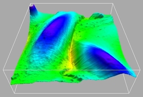 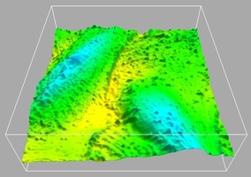
40 x 40 µm2 images before (left image) and after mannitol treatment (right image) rendered in 3D. Height scale 2.5 µm.
Related link: http://www.asylumresearch.com/CloseUp6.shtml
Reference:
Bálint Z., I.A. Krizbai, I. Wilhelm, A.E. Farkas, Á. Párducz, Z. Szegletes and G. Váró. 2006. Changes induced by hyperosmotic mannitol in cerebral endothelial cells: an atomic force microscopic study. European Biophysical Journal. In press.
| |