|
|
|
|
|
|
 | |
Photoelectric signals of membrane proteins |
Principal Investigators:
András Dér,
Lajos Keszthelyi,
Pál Ormos,
György Váró,
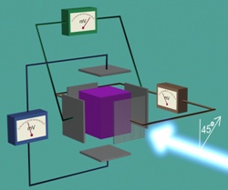
Due to the symmetry of the system the above method yields only one component of the charge displacement: the one in the
pumping direction, the membrane normal. Side motions average out in the sample. However, with photoselection it is
possible to determine motions in additional directions, too.
Photoselection: if a sample is partially excited with polarized light, an anisotropic excitation is achieved.
The ones in the direction primarily absorbing the light will be predominantly excited. This introduced ansitotropy permits
the detection of additional directional effects.
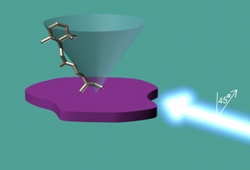
In the case of photoelectric experiments, however, photoselection is
not trivial: linear chomophores have no directionality that would be needed to see net charge motion (consider, if a
vertical absorber is selected, half point up, half down, and the coupled charge motions cancel out). In the case of the
oriented Bacteriorhodopsin/purple membrane there is a way, due to the oriented nature of the sample. All membranes point
in one direction, therefore the chromophores lie on the surface of a cone with axis normal to the membrane,
pointing in one direction. Illuminating this cone from the side with light of polarization making 45° with the cone axis
will effectively photoselect the molecules.
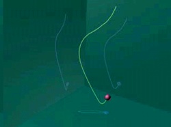
Now we can measure the charge motion in all three dimensions.
And we can determine the full trajectory of the charge motion. Click on the image below to play an animation.
References:
A. Der, L. Oroszi, A. Kulcsar, L. Zimanyi, R. Tóth-Boconádi, L. Keszthelyi, W. Stoeckenius, P. Ormos,
Interpretation of the spatial charge displacements in bacteriorhodopsin in terms of structural changes during
the photocycle, Proc. Natl. Acad. Sci. Usa 96 2776 (1999)
L. Oroszi, A. Der, P. Ormos, Theory of electric signals of membrane proteins in three dimensions,
European Biophysics Journal 31 (2) 136-144 (2002) View PDF
| |